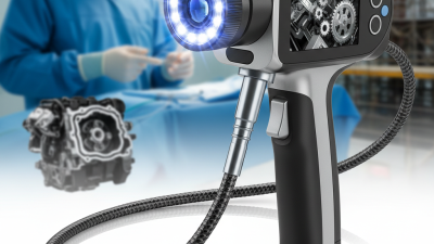

A Wireless Endoscope Camera is a compact imaging device that sends live video without a traditional cable connection. It combines a miniature camera, LED lights, a battery, and a wireless transmitter. The lens may be only a few millimeters wide, yet it can reveal narrow spaces with surprising clarity. A smartphone, tablet, or dedicated monitor receives the signal through Wi-Fi or another short-range connection.
Dr. Jacques Marescaux, a pioneer in minimally invasive surgery, has expressed a guiding principle for surgical technology: “Technology should assist the surgeon, not replace surgical judgment.” This idea remains important when examining a Wireless Endoscope Camera. Wireless transmission can improve movement around a workstation and reduce cable clutter. However, convenience does not guarantee clinical accuracy. Image lag, weak signals, glare, limited battery life, and lens fogging can affect what the operator sees.
The camera is only one part of the system. Its sensor captures light, software processes the image, and the transmitter sends the data to a receiving device. Some models offer high-definition video, adjustable lighting, image recording, or disposable protective covers. These features sound impressive. Real performance depends on testing.
A careful user should check image stability, cleaning requirements, operating distance, and data security. The device also needs appropriate training and professional supervision. It is not a magic tool. Its value comes from dependable images, thoughtful handling, and sound medical decisions. This article explains how the technology works, where it performs well, and where its limitations deserve honest attention.
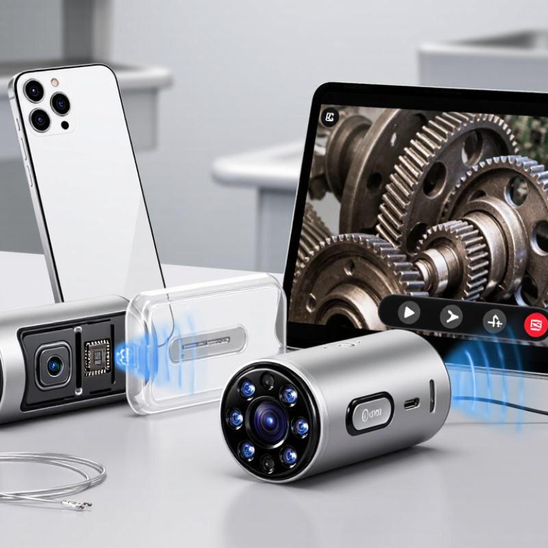
A wireless endoscope camera is a compact imaging device used to view narrow, enclosed, or difficult-to-reach spaces. Unlike a conventional wired scope, it sends captured images to a nearby screen or mobile device without a continuous video cable. The insertion tube still enters the inspection area. Wireless describes the data connection, not the entire instrument.
Its core begins with a miniature camera sensor and a small lens at the tip. Surrounding LED lights illuminate dark cavities and help reveal scratches, corrosion, moisture, or loose parts. A flexible insertion tube guides the camera through bends, while a handle provides directional control. Inside the handle, a processor converts the camera’s signal into digital images. A wireless module then transmits those images over a short-range network.
Power usually comes from a rechargeable battery. This makes movement easier, but battery life can limit long inspections. The receiving device may show live video, store photographs, or record short clips. Some systems include brightness controls, image rotation, and adjustable focus. These features sound simple. They are not always simple in use.
Image quality depends on lighting, lens cleanliness, distance, and wireless interference. A bright screen cannot correct a blurred lens. In practical work, users should check the battery, test transmission, and inspect the tube before use. Cleaning procedures also matter, especially when the camera enters dusty or damp environments. Wireless convenience improves access, yet it does not replace careful handling, suitable training, or professional judgment.
A wireless endoscope camera captures images through a tiny lens and sensor at its tip. Most modern systems use a CMOS image sensor, which converts incoming light into electrical signals. A miniature LED ring illuminates dark cavities, pipes, or equipment surfaces. Light comes first.
The camera processor then adjusts exposure, color, and sharpness before compressing each frame. It sends the data through Wi-Fi or another short-range wireless connection to a phone, tablet, or monitor. Cisco’s Annual Internet Report projected video would represent 82% of internet traffic, showing why efficient compression and stable bandwidth matter. However, image quality depends on more than resolution. Lens position, lighting, sensor size, interference, and distance can change what the operator actually sees. Small details matter.
Fortune Business Insights’ 2024 medical endoscopes report estimated the global market at about 28.7 billion U.S. dollars in 2023. That growth reflects rising demand for compact visualization tools, although wireless cameras are only one part of the market. In practical testing, a camera may advertise high definition yet produce blurred images during movement. Transmission delay can also make fine control difficult. A reliable design should balance resolution, frame rate, battery life, and connection stability. I have found that a steady 720p feed can be more useful than unstable ultra-high definition. That trade-off deserves more attention.
| System Component | Function | How It Works | Typical Technical Details |
|---|---|---|---|
| Camera Sensor | Captures the scene at the tip of the endoscope. | A CMOS image sensor converts incoming light into electrical signals and digital image data. | Commonly CMOS; image sizes may range from basic VGA to Full HD, depending on the model. |
| Lens Assembly | Focuses light onto the sensor and determines the viewing angle. | The miniature lens gathers reflected light from the inspection area and projects it onto the sensor. | Often uses a fixed-focus lens with a short working distance; viewing angles commonly fall between about 60° and 90°. |
| LED Illumination | Provides light inside dark or enclosed spaces. | Small LEDs positioned near the lens emit visible light, while the camera sensor records the illuminated surface. | Brightness is commonly adjustable; illumination is usually white LED light. |
| Image Processor | Prepares raw sensor data for transmission and viewing. | The processor adjusts color, exposure, sharpness, and compression before sending the image stream wirelessly. | May output JPEG still images and compressed video formats such as H.264 or similar formats. |
| Wireless Transmitter | Sends images and video from the camera to a nearby display device. | A built-in wireless module creates or joins a local Wi-Fi network, allowing a phone, tablet, or computer to receive the stream. | Usually operates over the 2.4 GHz Wi-Fi band; range is often approximately 5–15 meters indoors, depending on obstacles. |
| Mobile or Computer Display | Shows the live inspection image and may store photos or videos. | A compatible application or web interface receives the wireless stream and renders it on the device screen. | Recording capability depends on the operating system, application, storage space, and camera resolution. |
| Probe or Insertion Tube | Guides the camera into narrow or difficult-to-reach areas. | The flexible or semi-rigid tube carries the sensor, lens, LEDs, and electrical connections to the inspection location. | Common outside diameters include approximately 3.9–8 mm, although sizes vary widely by application. |
| Power Supply | Provides energy for the sensor, LEDs, processor, and wireless module. | A rechargeable lithium-ion or lithium-polymer battery commonly powers portable wireless units. | Typical operating time is about 60–120 minutes, depending on LED brightness, wireless activity, and battery capacity. |
| Protective Housing | Protects internal electronics from dust, moisture, and physical contact. | Seals and protective materials limit the entry of liquids and particles around the probe and camera head. | Some inspection probes are rated IP67 or higher, but the rating must be verified for the specific model and component. |
| Image Transmission Process | Moves captured visual information from the probe to the viewer. | Light becomes electrical data at the sensor, the processor compresses it, and the wireless transmitter sends packets to the receiving device. | A short delay is normal; latency increases when the signal is weak or the video resolution and network traffic are high. |
| Common Applications | Allows visual inspection without fully dismantling equipment or entering confined spaces. | Users guide the probe through openings to inspect pipes, engine areas, machinery, wall cavities, drains, and other restricted locations. | Useful for inspection and documentation; it is not automatically suitable for medical use or hazardous environments. |
Note: Specifications are representative ranges for wireless endoscope cameras. Actual performance varies by design, environment, wireless interference, and operating conditions.
A wireless endoscope camera combines a miniature lens, LED lights, an image sensor, and a radio transmitter. Unlike a wired model, it sends live images to a nearby phone or tablet. The camera usually creates a private Wi-Fi connection. Some systems use Bluetooth for pairing, but Wi-Fi carries video more effectively.
On a workbench, a technician can guide the flexible probe through a narrow pipe while watching the screen. This removes the need to lean over the opening.
This matters.
The mobile device acts as both monitor and control panel. An app may adjust brightness, capture photographs, record video, or rotate the view. Connection quality depends on distance, walls, radio interference, and battery level. When the signal weakens, the image can freeze or show a delay.
That delay is easy to underestimate. For inspection, even a few seconds may hide a moving leak or damaged edge. Secure pairing and encrypted transmission matter when images contain sensitive workplace or patient information. Users should follow privacy requirements and verify device compatibility before saving recordings.
From practical testing, lighting often matters more than advertised resolution. A bright reflection can hide a crack, while dust can soften detail. Clean the lens carefully. Wireless designs also require charging, and a dead battery stops the inspection immediately.
I would not treat a phone display as final evidence without checking the original file and viewing conditions. That limitation deserves attention.
For medical use, trained personnel must interpret findings and follow approved procedures. For technical work, important measurements should be confirmed with suitable tools.
Mobile access improves inspection speed, but it does not replace human judgment.
A wireless endoscope camera is a small inspection tool with a flexible cable, camera lens, and built-in light. It sends live images to a phone, tablet, or monitor through a wireless connection. This design helps users view narrow spaces without carrying a larger display. It can support equipment checks, household repairs, and professional inspections. Medical applications require approved equipment and trained professionals.
Charge the camera and install its official viewing application. Turn on the camera, then connect your device to its wireless signal. Keep the phone within a practical range, because walls and metal surfaces may weaken the connection. Check the image before inserting the cable. Adjust the light level until edges look clear, not washed out.
Guide the cable slowly through the target space. Use gentle movements, and avoid forcing the tip around sharp bends. Watch the live screen while rotating the handle. You can capture photos or short videos for later review. A common mistake is moving too quickly. Pause when the image shakes. Clean and disinfect the camera according to its instructions after use. Never place an unapproved camera inside a person or animal. I have found that a second viewing angle often changes the initial judgment, so inspect uncertain areas again before making repairs.
A wireless endoscope camera combines a miniature lens, LED lighting, image sensor, transmitter, and rechargeable battery. It sends live video to a phone, tablet, or monitor without a fixed video cable. In practice, technicians use it inside pipes, engines, wall cavities, and narrow machinery gaps. Medical teams may use similar systems for visualization, but clinical use requires professional training, validated equipment, and strict infection-control procedures.
Its main benefit is access. A flexible probe can reach around bends while the operator watches a bright image nearby. This can reduce disassembly time and support faster maintenance decisions. Grand View Research estimated the global medical endoscopy market at about USD 30.3 billion in 2023, showing strong demand for minimally invasive visualization tools. However, a wireless camera is not automatically precise. Signal loss, battery limits, glare, moisture, and low light can distort important details. Small screens may also hide cracks or tissue changes. The operator can feel confident too quickly. That remains a real weakness.
Tips: Test the connection before entering a confined space. Keep a spare battery available. Clean the probe according to its approved instructions. Record close-up images and wider context. Never treat a camera image as a final diagnosis or structural safety decision without qualified review. A 2024 FDA guidance update also stresses appropriate reprocessing and infection-control practices for reusable endoscopic devices.